A rare case of pulmonary sequestration associated with a double cardiac shunt has been reported in a 6-month-old female infant in Cameroon. The infant, born at term following a singleton pregnancy, was delivered vaginally and was the only child of a teenage couple. She was seen at the paediatric cardiology clinic at the Abidjan Institute of Cardiology for recurrent rhinobronchitis and failure to thrive in terms of height and weight, which had been present since she was 3 months old.
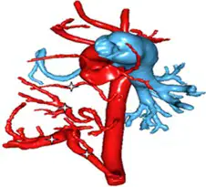
Clinical and diagnostic investigations revealed pulmonary sequestration of the right lower lobe associated with dextrocardia, an atrial septal defect, and patent ductus arteriosus complicated by pulmonary arterial hypertension. The infant's condition was further complicated by a right subclavian artery lying behind the oesophagus. The diagnosis was made using multimodal cardiovascular imaging, which is essential to guide the therapeutic strategy for such complex cardiopulmonary malformations.
Pulmonary sequestration is a rare congenital malformation of the lung tissue, which may be associated with other cardiac abnormalities. The condition occurs when a portion of lung tissue receives its blood supply from an anomalous systemic artery rather than the pulmonary artery. This can lead to various complications, including respiratory distress, heart failure, and pulmonary hypertension.
The infant's condition was managed by proposing treatment abroad due to the complexity of the cardiopulmonary malformation and the limitations of the local facilities. The therapeutic strategy for pulmonary sequestration associated with cardiac shunts typically involves surgical or transcatheter intervention to close the shunt and remove the sequestered lung tissue. In some cases, arterial embolization may be used to occlude the anomalous systemic artery supplying the sequestered lung tissue.
Pulmonary sequestration may be associated with various congenital heart defects, including atrial septal defects, patent ductus arteriosus, and ventricular septal defects. A review of the literature revealed that multimodal cardiovascular imaging is crucial in diagnosing and managing such complex cardiopulmonary malformations. The use of imaging modalities such as echocardiography, computed tomography, and magnetic resonance imaging can help guide the therapeutic strategy.
The case report highlights the importance of considering pulmonary sequestration in the differential diagnosis of infants presenting with recurrent respiratory symptoms and failure to thrive. Early diagnosis and treatment can significantly improve outcomes in infants with pulmonary sequestration associated with cardiac shunts. A multidisciplinary approach involving paediatric cardiologists, cardiothoracic surgeons, and radiologists is essential in managing such complex cases.
The study was published in the Health Sciences and Diseases journal, a peer-reviewed publication that aims to disseminate knowledge on various aspects of health sciences. The authors of the study emphasized the need for further research on pulmonary sequestration associated with cardiac shunts to improve our understanding of this rare congenital condition.
Key points
- - Pulmonary sequestration is a rare congenital malformation of the lung tissue that may be associated with other cardiac abnormalities. - Multimodal cardiovascular imaging is essential to guide the therapeutic strategy for complex cardiopulmonary malformations. - Early diagnosis and treatment can significantly improve outcomes in infants with pulmonary sequestration associated with cardiac shunts.