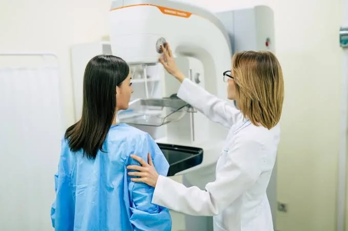
A new artificial intelligence system that combines breast ultrasound and digital breast tomosynthesis has shown promising results in improving cancer detection while reducing false-positive rates. The model, trained on images from 2,187 breasts, was evaluated on two independent test sets of 632 and 500 breasts verified by tissue biopsy. This innovative approach aims to support clinicians in distinguishing malignant from benign findings, particularly in dense-breasted women.
The AI model was tested in six different configurations, including three single-modality models using ultrasound, digital mammography, and DBT, and three dual-modality models pairing each scan type with another. The ultrasound-DBT hybrid model outperformed all others, achieving a 95.5% specificity for non-cancerous lesions while maintaining an 85% sensitivity for cancer detection. This significant improvement in performance highlights the potential benefits of combining multiple imaging modalities.
The model's strength lies in its ability to reduce false-positive results without compromising its ability to detect cancer, especially in dense-breasted women and for lesions under two centimeters. By merging ultrasound's detailed information on lump morphology with DBT's three-dimensional structural view, the AI system provides radiologists with a clearer decision-making aid. This enhanced diagnostic accuracy can lead to better patient outcomes and reduced healthcare costs.
The researchers emphasize that the AI tool is intended to support, not replace, radiologists in their clinical decision-making. The current validation of the model is limited to a single center and retrospective data, highlighting the need for larger, multi-center trials to confirm its performance across diverse equipment and patient populations. These future studies will be crucial in determining the model's readiness for routine clinical adoption.
The development of this AI model is a significant step forward in the use of artificial intelligence in breast cancer diagnosis. By leveraging the strengths of multiple imaging modalities, the model has the potential to improve diagnostic accuracy and reduce the burden on healthcare systems. Further research is needed to fully realize the benefits of this technology and to address any challenges that may arise during its implementation.
The study's findings have important implications for breast cancer diagnosis, particularly in Kenya and other countries where access to advanced imaging technologies may be limited. The use of AI-powered diagnostic tools could help bridge the gap in healthcare disparities and improve health outcomes for patients in these regions. As the model continues to evolve, it may also be adapted for use in other medical applications.
In conclusion, the AI model combining ultrasound and 3D mammography has shown promising results in improving breast cancer detection and reducing false positives. While further research is needed to confirm its performance, this innovative approach has the potential to make a significant impact on breast cancer diagnosis and treatment.
Key points
- The AI model achieved a 95.5% specificity for non-cancerous lesions while maintaining an 85% sensitivity for cancer detection.
- The model's strength lies in reducing false-positive results without compromising its ability to spot cancer, especially in dense-breasted women and for lesions under two centimeters.
- The AI tool is intended to support, not replace, radiologists in their clinical decision-making.